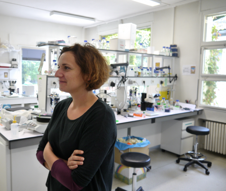
Meet our speakers
-

Letizia Fontana
-
Benjamin Lepennetier
-

Florence Assan
-

Helena Kissiova
Abstracts Conference part 1
-
Base Editing of BCL11A cis-regulatory Regions to Treat β-hemoglobinopathies
Abstract : Sickle cell disease (SCD) is caused by a mutation affecting adult hemoglobin (Hb) expression and causing red blood cell (RBC) sickling. Transplantation of autologous genetically modified hematopoietic stem cells (HSCs) is a therapeutic option. The clinical severity of SCD is alleviated by fetal Hb (HbF) reactivation, which can be achieved by down-regulating BCL11A, a major HbF repressor. To reduce BCL11A levels, CRISPR/Cas9 approaches targeting erythroid-specific BCL11A enhancer have been recently approved for treatment of β-hemoglobinopathies. However, genotoxicity concerns arise from CRISPR/Cas9-induced double-strand breaks (DSBs). To overcome this limitation, we exploited DSB-low/free base editors (BE) to introduce point mutations within the binding sites (BS) of GATA1 and ATF4 transcriptional activators in the BCL11A enhancers and reduce BCL11A expression. We generated different nucleotides changes within the BSs in SCD-derived HSCs, with an overall editing efficiency that reached up to 90%. Despite the fact that HbF reactivation was observed in all edited samples, the HbF levels were not sufficient to achieve the full rescue of the sickling phenotype in RBCs. Hence, we decided to simultaneously target the GATA1 and ATF4 BS with the aim of further increasing HbF levels. Erythroid cells differentiated from SCD HSCs edited at both GATA1 and ATF4 BS showed high HbF expression (representing up to 34% of the total Hb) and exceeding the levels achieved using the CRISPR/Cas9 nuclease-based strategy. Finally, HbF reactivation was sufficient to allow a substantial rescue of the sickling phenotype, decreasing the proportion of sickling cells to only 16%. In conclusion, we have provided evidence of the effectiveness of simultaneous base editing of HbF trans-regulatory elements for correcting the sickling phenotype. This proof-of-concept work will enable the pre-clinical and clinical development of base-edited HSCs for the therapy of β-hemoglobinopathies.
-
Decrypting the impact of defective PRDM13 function on hindbrain development using zebrafish and organoid models
Abstract : Developmental disorders of the cerebellum (DDC) form a heterogeneous group of neurodevelopmental disorders. Surprisingly, mutations in key developmental transcription factors have been rarely described as causative in DDC. We recently identified homozygous mutations in PRDM13 in children presenting severe and early-onset hypoplasia of the cerebellum and inferior olivary nucleus (ION), and a reduction and disorganization of the Purkinje cell (PC) layer. PRDM13 encodes a transcriptional repressor, critical for neural cell fate specification in the retina and spinal cord. Its function in hindbrain development was still unknown. To validate the role of PRDM13 in the pathology and the underlying mechanisms, we analyzed prdm13 expression and function in zebrafish. We show that prdm13 is expressed in specific progenitors in the cerebellum and posterior hindbrain, as in human embryonic samples. Lineage-tracing shows that in the posterior hindbrain prdm13+ progenitors directly give rise to multiple dorsal neuronal populations and to the ION. In the cerebellum, their progeny comprises mainly PCs and GABAergic interneurons. In prdm13 zebrafish mutants, we observe profound fate specification defects in the posterior hindbrain, with an absence of the ION and inhibitory neuronal populations contributing to viscerosensory columns. In the cerebellum, the phenotype is less severe, with a reduction of a specific PCs subpopulation. By RNA sequencing in prdm13+ cells, we confirm the deregulation of multiple fate specification genes and unravel an additional impact on proliferation. Strikingly, this approach also highlights a leaky expression of pancreatic genes in mutant cells. Altogether, our work demonstrated the causative role of PRDM13 variants in affected children and its key roles in hindbrain development. We are now generating human cerebellar-like organoids carrying PRDM13 mutations to complement the observations in zebrafish and further decrypt the pathological mechanisms.
-
Pathogenic loss-of-function mutations in ADAR1 in severe psoriasis drive an interferon signature
Abstract : Psoriasis (PSO) is an inflammatory skin disease affecting 5% of population. Monogenic models of PSO remain rare to date. Adenosine Deaminase Acting on RNA 1 (ADAR1) is an A/I editing enzyme, which loss-of-function (LOF) mutants account for Aicardi-Goutières syndrome (AGS) interferonopathy. We report the association of ADAR1 variants with early onset PSO.
Whole exome sequencing (WES) identified in a family with autosomal dominant early onset PSO and interferonopathy-like phenotype in proband, a heterozygous (htz) missense mutation in the activity domain of ADAR1 (c.3355G>A) co-segregating with PSO, type I interferon (IFN) signature in blood, and a decrease of ADAR1 protein expression in patients’ keratinocytes (kerat.).
WES in a cohort of 115 PSO patients identified 11 other ADAR1 htz variants in severe PSO patients, presenting a specific response to JAK1 inhibitor. ADAR1mut PSO patients carried a significant increase of blood IFN-score vs. ADAR1wt PSO patients, bulk transcriptomic studies in PSO skin showed an enrichment of IFN-associated terms. In PSO skin, single cell transcriptomic studies showed the role of kerat. in expressing ISG, immunostaining showed a keratinocyte activation of pSTAT1.
To assess the impact of ADAR1 loss in primary kerat., siRNA knock-down (KD) was performed. ADAR1KD was associated in transcriptomic studies with a significant increase of ISG and some PSO-related genes (CXCL8, IL23A, CCL20), an enrichment of IFN-related terms. Mass spectrometry revealed an enrichment of IFN-related terms (confirmed in WB with an activation of pSTAT1) and a down-regulation of proteins involved in cornification. RNAseq editome showed a drastic reduction (84.4%) of A/I editing in alu sequences. Human skin equivalent using ADAR1KD cells presented an alteration of the skin structure.
Altogether, this establishes a new single gene model of PSO due to ADAR1 LOF mutations, driving IFN signature with a kerat. scenario, and opening new avenues for precision medicine.
-
RCD2 and Bortezomib – a story yet to be told?
Abstract : Celiac disease (CeD) is an immune-mediated enteropathy triggered by dietary gluten. A rare complication is the onset of invasive enteropathy-associated lymphomas (EATL), often preceded by low-grade lymphoproliferation called type II refractory CeD (RCD2). Recent findings showed recurrent JAK1/STAT3 gain-of-function mutations in all lymphomas complicating CeD. However, RCD2 cells have high intra-tumor heterogeneity that could foster resistance to JAK/STAT-targeted therapy. Considering Bortezomib's (BTZ) clinical success in hematological disorders, we have explored its effects on RCD2 tumor cells. In vitro studies using BTZ-treated RCD2 cell lines selectively eliminate malignant RCD2 cells while preserving normal T cells, suggesting no interference with the immune anti-tumor response. To further investigate BTZ as a therapeutic option for RCD2, we have developed two complementary models. First, an air-liquid interface organoids model using fresh intestinal biopsies allows studying BTZ's effect on RCD2 cells and its microenvironment. Preliminary results show successful cultivation of gut epithelium cell populations for up to 10 days. scRNAseq on BTZ-treated RCD2 organoids will be performed to assess its impact. Second, we have established a xenograft model using RCD2 cells lines transferred in immunodeficient mice to assess the effects of BTZ on survival and tumor infiltration through in vivo imaging and flow cytometry. Finally, positive clinical response in two RCD2 patients treated with BZM was observed. sRNAseq on blood and duodenal biopsies of one patient pre and post BZM treatment will define functional signatures of key signaling pathways. This study will help to elucidate BTZ efficacy reasons and propose it as a promising option for RCD2 patients, given its selective tumor killing potential.
-

Rosalie Borry
-

Pierre Duquesne
-

Nicole Bertola
-

Fokion Spanos
Abstracts Conference part 2
-
ALK-induced lymphomagenesis: from CRISPR modeling to the identification of new regulators by OMICs analyses
Abstract : ALK+ anaplastic large cell lymphoma (ALCL) is a rare pediatric lymphoma harboring most frequently the t(2;5)(p23;q35) chromosomal translocation resulting in NPM1-ALK fusion protein. The first-line treatment is polychemotherapy which yields a high response rate but also presents severe side effects with 30% of relapses of poor prognosis. Second-line treatments now include targeted therapies, underscoring the need for a better comprehension of the pathology. In our team, we have developed a model of ALK+ ALCL transformation. Using CRISPR, we can induce the t(2;5)(p23;q35) translocation and reproduce ALK+ ALCL oncogenesis from healthy primary T lymphocytes to the tumors. Importantly, the edited tumors faithfully recapitulate the immune and histological phenotypes of patients. This original and robust model offers an extensive time frame to analyze every crucial stage of cell transformation, from the cell of origin to in vivo tumors, including pre-transformed cells. To obtain a comprehensive understanding of cell transformation, we are integrating OMIC data including transcriptomics (bulk and scRNAseq), epigenetics (ATAC-seq), and replication timing (Repli-seq). We have recently identified new candidate regulatory genes of ALK+ driven oncogenesis and are currently in the process of functionally validating them. Besides the t(2;5)(p23;q35) translocation, novel somatic mutations in patient tumors have recently been identified by a collaborator, however their exact role in oncogenesis remains unclear. To decipher their function, we are using a CRISPR KO approach in our model and already demonstrated a strong alteration of the transcriptomic oncogene signature. This includes the upregulation of several pathways, including the Hippo pathway involved in cell proliferation and apoptosis. Furthermore, these mutations strongly increased the migration and invasion potentials of cells. We are now conducting mouse experiments to assess the in vivo significance of these mutations.
-
Description of the novel role of kinesin-1 in dendritic cell migration : a bridge between microtubular dynamics and actin pools.
Abstract : Dendritic cell (DC) migration is a complex process crucial for the establishment, maintenance and development of immune responses. Immature DCs migrate using a biphasic migration pattern, alternating between a slow phase, necessary for the sampling of the antigen and the scanning of the immediate cell environment, and a fast phase, essential for the motility of the cell. While extensive research has been done on understanding DC migration and its necessity in the setting-up of immune responses in patients, the role of kinesin-1, a molecular motor using the microtubular network, in this process remains unclear. We previously showed that kinesin-1, is a key regulator in antitumoral responses and cross-presentation, a specific antigen presentation mechanism in DCs. Nevertheless nothing has yet been established about its implication regarding DC migration. In this study, we investigated a novel role of kinesin-1 on DC migration, specifically during the establishment of the slow phase of their migratory behavior. Indeed, kinesin-1 deficient DCs display an erratic migratory behavior, lacking this slow phase, as well as an altered cell plasticity. Through live-cell imaging, microfluidics and genetic knockdown approaches, we discovered that kinesin-1 regulates the actin cytoskeleton dynamics, known for its essential role in the migratory behavior of DCs and in macropinocytosis, the engulfment of extracellular material. We hereby suggest that kinesin-1 acts as a cross-linker between microtubules and actin pools. Furthermore, our latest results, associated to drug testing, suggest that impairment of kinesin-1-related transport across the microtubular network could lead to the disruption of molecular pathways essential for the maintenance of the cytoskeletal dynamics. Thus, our work contributes to a better understanding of DC migration, and highlights the complex interplay between kinesin-1, actin dynamics, microtubular network, and its role on immune response modulation.
-
De novo mutations in the ATOH1 transcription factor induce a characteristic brainstem malformation
Abstract : Developmental disorders of the cerebellum (DDC) represent a heterogeneous group of diseases whose underlying mechanisms remain often unclear. Our recent works highlight early deregulations in the transcriptional control of neuronal fate specification as a pathological process that could contribute to a significant number of DDC cases. In our cohort of patients, we identified de novo variants in genes encoding key transcriptional regulators. Among those, we identified two de novo variants in ATOH1, associated with similar clinical presentations: cerebellar hypoplasia, a specific malformation of the pons, motor and cognitive impairments and deafness. In animal models, ATOH1 activity was shown to be necessary for neural fate specification in the cerebellum and brainstem and for mechanosensory hair cell differentiation, making ATOH1 a strong candidate causative gene for these patients. The two identified mutations lead to a frameshift, and thereby to a truncation of the C-terminal domain of ATOH1. Alterations of this domain have been associated with an increase in the stability of ATOH1 protein and we thus suspect unusual gain-of-function (GOF) effects of these de novo variants. Indeed, our in vitro results show an increased protein stability compared to the WT form, thus reinforcing our hypothesis. In parallel, we are investigating the variant’s transcriptional activity compared to the WT protein. We are also assessing in vivo the effect of truncating mutations in the C-terminal domain using a CRISPR/Cas9-based zebrafish model. Here, we observed a transient increase in the number of mechanosensory hair cells in the neuromasts of the lateral line at 48hpf, corroborating the hypothesis of a GOF and representing a possible link with the deafness of the patients. In the hindbrain, our preliminary results point to a premature differentiation of the neuronal population derived from atoh1a-expressing progenitors, which might be responsible for the brainstem malformation.
-
Interaction between intestinal bacterial and human amyloids in Parkinson’s disease
Abstract: Emerging evidence suggests Parkinson's disease (PD) may originate in the gut, with α-synuclein (A-SYN) aggregates found in the gastrointestinal tract prior to the onset of motor symptoms. Functional bacterial amyloids (FuBAs), notably Curli, accelerate PD progression by promoting A-SYN aggregation in experimental models. However, the precise mechanisms underlying PD pathology in the gut remain elusive. We hypothesize that enteroendocrine cells (EECs), expressing A-SYN and exposed to intestinal content, play a crucial role by internalizing FuBAs into lysosomes, fostering A-SYN aggregation and transmission to enteric neurons. We studied Curli/A-SYN interaction using the EEC line HuTu80 and human induced pluripotent stem cell (hiPSC)-derived enteric neurons and intestinal organoids (HIOs). CsgA-WT (monomers constituting curli), CsgA-R12343 (non-amyloidogenic control), and Curli were purified and characterized by thioflavin-T assay and electron microscopy. HuTu80 were treated with CsgA-WT, CsgA-R12343, or Curli. We found that CsgA monomers and Curli were localized into lysosomes in HuTu80. Furthermore, CsgA-WT, but not CsgA-R12343, is degraded in lysosomes as shown by bafilomycin A1 treatment. Preliminary data indicate that CsgA-WT and Curli induce A-SYN accumulation in HuTu80 co-treated with exogenous A-SYN monomers. The molecular mechanisms of Curli/A-SYN cross-seeding will be investigated by whole-cell and lysosome proteomics. Lysosomes are immunoprecipitated using HuTu80 stably expressing a tagged lysosomal membrane protein (TMEM192-3xHA). To study cross-seeded amyloid transfer between EECs and neurons, we established a microfluidic co-culture of hIPSC-derived enteric-like neurons and EECs. In summary, our results highlight the role of lysosomes in amyloid cross-seeding. Using HIOs co-cultured with enteric neurons, we now aim to validate our results in a disease-relevant model and identify pathways targeting early disease stage.
-

Amaia Ochandorena-Saa
-

Federico Bertoli
-

Patricia Panikulam
Abstracts Conference part 3
-
Mechanisms of heterotaxy and situs inversus in a mouse model of motile ciliopathy
Abstract : The mammalian heart, which functions as a double left-right pump, depends on the left-right patterning of the embryo. Asymmetry emerges in the left-right organiser or node, in which motile cilia create a fluid flow towards the left. This induces the left-sided expression of the secreted factor NODAL, which is essential to pattern heart precursors and shape the tubular heart primordium into an asymmetric rightward helix or loop. Inactivation of Nodal leads to the heterotaxy syndrome with right isomerism of lungs and atria and with complex congenital heart defects. Although heterotaxy is often described as a random process, situs inversus is never observed in Nodal mutants. In this project we targeted left-right asymmetry upstream of Nodal, and used Ccdc40 mutant mice as a model to impair motile cilia of the node. Ccdc40 mutant mice show a different phenotype compared to Nodal mutants, including situs inversus and heterotaxy with left isomerism. Earlier in development, Ccdc40 mutants show inverted heart looping with partial penetrance. By quantifying morphological anomalies and using genetic tracing of NODAL signalling, we have identified four variants of heart looping, potentially reflecting the four categories seen at birth – unaffected individuals, situs inversus, heterotaxy with rightward looping and heterotaxy with inverted looping. Our ongoing work includes a transcriptomic analysis of left and right heart precursors, to uncover the asymmetric gene expression signature of the different phenotypic outcomes. In addition, we have optimized a protocol to measure perturbations of node flow in live Ccdc40 mutant embryos. This work provides novel insights into the mechanisms of asymmetric heart morphogenesis and is relevant to complex congenital heart defects.
-
Investigating Parkinson’s Disease-associated Alpha-Synuclein Pathology in Midbrain Organoids
Abstract : Parkinson’s disease (PD), the most common age-related degenerative movement disorder, is characterized by the progressive loss of dopaminergic neurons in the substantia nigra pars compacta, leading to motor impairment and non-motor symptoms. A key feature of PD pathology observed in most patients is the aggregation and spread of misfolded alpha-synuclein (α-syn). However, the causal relationship between α-syn pathology and PD remains unclear. This lack of knowledge is largely due to the limitations of current PD research models, including 2D in vitro cellular models and animal models. These models fail to faithfully recapitulate the intricate α-syn pathology observed in the human brain, thus hindering the clinical translation of research findings. Here, we developed induced pluripotent stem cell (iPSC)-derived midbrain organoids as a more advanced and accurate model to study the dynamics and mechanisms of α-syn pathology in PD. To model idiopathic PD, we induced endogenous α-syn aggregation in healthy control-derived organoids using exogenous α-syn preformed fibrils (PFFs). PFF-treated organoids showed neuronal internalization of α-syn fibrils and α-syn pathology over time. Interestingly, single-cell RNA sequencing revealed a dysregulation of oxidative phosphorylation in PFF-treated organoids. Moreover, using iPSC-derived midbrain organoids integrating isogenic microglia, we are currently investigating the role of microglia in α-syn pathology. Our study provides an advanced modeling approach that will contribute to the identification of the role of α-syn and glial cells in PD.
-
Role of PI4KIIIa in cell migration
Abstract: Actin related primary immunodeficiency disorders (PIDs) or actinopathies is a group of primary immunodeficiency disorders characterised by mutations in actin or actin regulatory genes. Mutations in PI4KA gene in humans have multiple consequences ranging from leukodystrophy to immunodeficiency. We have a cohort of 5 patients with variants in PI4KA gene. PI4KA codes for the kinase PI4KIIIa that catalyses PI to PI4P. PI4P is the precursor of different phophoinositides (PIPs) which are involved in many basic cellular activities. PI4KIIIa is stable only when it forms a complex with its binding partners i.e., TTC7A/B, EFR3 and FAM126. Recent studies in the laboratory have shown that defects in TTC7A can alter actin dynamics, cell migration and survivability. Therefore, we speculate that defects in PI4KIIIa will also manifest similar cellular consequences. Preliminary results showed that 3 of the 5 patients have reduced amount of PI4KIIIa protein, suggesting that these variants could be deleterious in nature. In physiology, immune cells experience different kinds of constraints due to environmental characteristics. Using microfluidics, we mimic some of these restraints on cells in vitro to study different aspects of amoeboid cell migration, chemokine response and nuclear deformation capacity. We observed that some of the patient derived T cell blasts and B-LCL cells migrated faster in 1D microdevices as compared to control cells. One of these fast-moving patient cells also had a larger pool of actin at the rear end of the cell as opposed to the uniform distribution of actin in the control cells. All these suggest that PI4KIIIa is an indirect regulator of actin cytoskeleton dynamics and cell motility. In this study we wish to characterize the deleterious nature of PI4KA variants and the molecular mechanisms through which PI4KIIIa regulate actin dynamics.
-

Marta Martin Corredera
-

Hassan Saei
-

Alexis Bertrand
-

Maxime Heintzé
Abstracts Conference part 4
-
Feeder-free culture system for ex vivo generation of large numbers of NK and CAR-NK cells from hematopoietic stem and progenitor cells
Abstract: Natural Killer (NK) cells are currently under clinical investigation as an attractive alternative for immunotherapies since they neither cause graft-versus-host disease nor induce cytokine release syndrome. However, the use of these cells is restrained by the limited availability of ex vivo clinical-grade methods for their expansion and genetic modification. We patented a 7-day feeder cell-free culture system for ex vivo generation of ProTcell™ from CD34+ cells. This culture system can be combined with lentiviral vector transduction, generating up to 70% of transduced ProTcellTM. Here, we exploited the dual differentiation potential (T/NK) of ProTcell™ to develop an efficient method for producing large numbers of NK cells. ProTcell™ were cultured between 7 and 14 days in NK differentiation culture conditions (SCF/FLT3-L/IL-2/IL-7/IL-15). Large numbers (up to 10,000 NK cells per CD34+ cell seeded at day 0) of highly pure NK cells (>90% CD3-CD56+) were generated. These cells expressed activating receptors (NCRs, NKG2D, DNAM-1), NK transcription factors (Eomes, T-bet, ID2), and did not express inhibitory KIRs. They underwent degranulation and expressed IFNγ, TNFα, Perforin, and Granzyme B upon stimulation with K562 cells. When transduced with an anti-CD19 CAR lentiviral vector, efficient expression of the CAR led to the killing of NALM-6 cells efficiently (70% of cytotoxicity for transduced NK as compared to 2.5% for non-transduced NK). Therefore, the unique combination of our short (7-day) ProTcell™ culture system and NK cell differentiation/expansion culture (7 to 14 days) provides a valuable and clinically applicable method to generate abundant pure NK and CAR NK cell populations. The engraftment potential, persistence and anti-tumor activity of generated NK and CAR-NK cells are currently under investigation in an NSG murine model transgenic for hIL-15.
-
X-linked Alport Syndrome: From transcriptomic diagnosis to preclinical assessment of splice-switching oligonucleotide therapy using patient-derived cells and kidney organoids
Abstract: X-linked Alport syndrome (XLAS) is a hereditary glomerulopathy arising from genetic mutations in the COL4A5 gene, encoding the α5 chain of the collagen IV [α5(IV)]. In the presence of α3(IV) and α4(IV) chains, encoded by COL4A3 and COL4A4, respectively, the α5(IV) chain forms heterotrimers crucial for the structure and function of the glomerular basement membrane (GBM), a key component of the glomerular filtration barrier. We have developed a targeted RNA sequencing approach to detect missing splicing variants in XLAS patients. Given that intronic variations impacting splicing can be targeted using splice-switching antisense oligonucleotides (ASOs), the development of robust in vitro models is essential for rapidly and effectively assessing disease molecular mechanisms and testing therapeutic options. We detected pathogenic splicing events in a cohort of 19 patients with a typical XLAS phenotype but without any pathogenic variant in the coding sequences. Having successfully identified and characterized the pathogenic variants, we designed ASOs and developed induced pluripotent stem cells (iPSC)-derived XLAS kidney organoids as a robust in vitro model for disease characterization and effective evaluation of ASO therapy. This model was characterized using single cell RNA sequencing and immunostaining of the basement membrane. Subsequently, to restore the expression of COL4A5 in both patient-derived fibroblasts and iPSC-derived kidney organoids, we applied ASO therapy and successfully restored canonical splicing in these models. Characterization of the XLAS kidney organoids confirms the relevance of in vitro organoid model for variant characterization and therapeutic approach evaluation.
-
Somatic genetic rescue in Shwachman Diamond syndrome
Abstract : Shwachman Diamond syndrome (SDS) is a rare genetic ribosomopathy leading to impaired protein synthesis, which causes numerous symptoms including bone marrow failure and neutropenia that can evolve to myelodysplasia syndrome or AML. Biallelic mutations in the SBDS gene are responsible of above 90% of the SDS cases and we recently identified biallelic EFL1 mutations as a novel cause of SDS. SBDS together with EFL1 remove the anti-association factor eIF6 from the pre60S ribosomal subunit, allowing its interaction with the 40S subunit to form the mature ribosome 80S. Natural acquisition of somatic genetic events over time participates to age-related diseases. However, in Mendelian diseases these events can, in rare case, counteract the deleterious effect of the germline mutation and provide a selective advantage to the somatically modified cells, a phenomenon dubbed Somatic Genetic Rescue (SGR). We recently showed that several somatic genetic events affecting the expression or function of eIF6 are frequently detected in blood clones from SDS patients but not in healthy individuals, suggesting a mechanism of SGR. While most of these somatic mutations induce eIF6 destabilization or EIF6 haploinsufficiency, one recurrent mutation (N106S) did not affect the expression of eIF6 but rather impact its ability to interact with the 60S subunit. In order to further investigate the functional consequences of EIF6 haploinsufficiency and N106S mutation in a context of SDS, I introduced via CRISPR/Cas9 these mutations in immortalized SDS patients’ and control cells. These original cellular models will allow to precisely determine their effect on several aspects of cellular fitness, including ribosome biogenesis, translation rate, and cellular proliferation. I will present the most recent results obtained from these models. Overall, this study will help to characterize how somatic eIF6 haploinsufficiency and N106S mutation confer a selective advantage in SBDS or EFL1 deficient cells.
-
Role of somatic mutations and chromoplexy in Ewing sarcomagenesis
Abstract: Ewing sarcoma (EwS) is an aggressive bone cancer primarily affecting children and young adults. The survival rate is low: 62% at 5 years. This cancer is characterized by a chromosomal translocation fusing the EWSR1 gene and one of the ETS transcription factor family member, most often FLI1 (85%) and ERG (10%). The resulting fusion proteins act as aberrant transcription factors. Additional mutations are found in patient tumors in STAG2 (member of the cohesin complex), TP53 and CDKN2A genes. In particular STAG2 plays a role in the regulation of the fusion oncogene transcriptional activity. Chromoplectic karyotype is also a hallmark of EwS tumors (found in 42% of patients). It consists in one burst of multiple non-reciprocal inter-chromosomal translocations, involving the one forming the fusion oncogene.
Our hypothesis is that the formation of translocation (EWSR1-ETS) is necessary but not sufficient to induce full tumorigenesis and has to be associated to genomic/genetic event(s) to regulate the toxicity of the fusion oncogene. This regulation could be favored by somatic mutations in STAG2, TP53 and CDKN2A and associated to chromoplexy.
Methods: By starting from mesenchymal stem cells (MSCs) derived from the bone marrow of EwS or healthy patients, we induce the specific EWSR1-ERG translocation with additional mutations by CRISPR/Cas9 systems. This translocation is always associated to chromoplexy in patient tumors. We strongly increase translocation events in our cells by using inhibitors of DNA repair pathways. Positive cells for EWSR1-ERG are then analyzed on a genomic level (translocations, chromoplexy).
Results: We managed to successfully recapitulate the transformation steps from EWSR1-ERG engineered MSCs by adding combination of somatic mutations. We are extensively characterizing (genome analysis, transcriptomic analysis…) these new models of sarcomagenesis to decipher the role of these mutations and genome instability in EwS transformation.
Scientific jury
